
Osteoarthritis and calcification
")
Cartilage calcification in OA
Osteoarthritis (OA) is characterized by progressive cartilage degradation, new bone formation and biomechanical deterioration of the joint. Cartilage damage leads to joint space narrowing, resulting in pain and loss of function. The disease is common, and was previously attributed to ageing and wear and tear. Calcium crystals are found in the joint fluid, cartilage and synovial membrane of OA joints, and correlate with the severity of OA.
The most common crystals are composed of basic calcium phosphate (BCP or hydroxyapatite), followed by calcium pyrophosphate (CPP). They are found in the cartilage, synovial fluid and synovial membrane. Analyses of joint cartilage revealed BCP crystals in 100% of OA samples, and CPPD crystals in 20%. This severity of calcification was positively correlated with OA severity, suggesting that calcification plays a role in the progression of OA.
Mechanisms of cartilage calcification
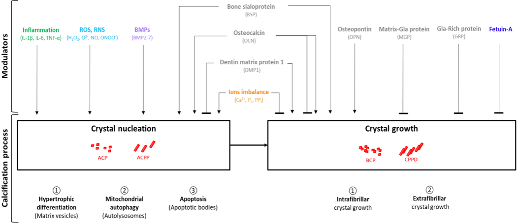
Cartilage cells (chondrocytes) are capable of forming calcium crystals normally and this process is enhanced when chondrocytes age, or when they are subjected to stresses such as inflammation, cell injury or cell death. The process starts with the appearance of microscopic aggregates in cellular vesicles (crystal nucleation in the figure) and ends with the formation of organised crystalline deposits (crystal growth) outside cells, bound to collagen and other matrix components. Multiple factors (inflammatory mediators, growth factors, ionic balance) play a part in this, as illustrated by the figure above.
Once formed, crystals can induce local inflammation via the secretion of inflammatory cytokines such as IL1ß and IL1a, IL6 which amplify inflammation driven cellular pathways. Crystals also cause oxidative stress and promote apoptosis (programmed cell death) as well as necrosis. The removal of calcium crystals once deposited in tissues is extremely difficult.
Synovial membrane calcification
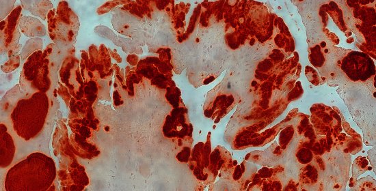
Calcium deposits are commonly found in the synovial membrane in OA, though they are not routinely searched. The photograph shows extensive red staining deposits (Alizarin red staining) in a patient with advanced OA who required a joint replacement.